Advanced Diffusion MRI in Renal Cancer: Oncologic Control and Renal Functional Reserve

Overview

In this project we investigate advanced diffusion MRI methods for the diagnosis of kidney tumors, estimation of the glomerular filtration rate, and prognosis of renal function after surgery.

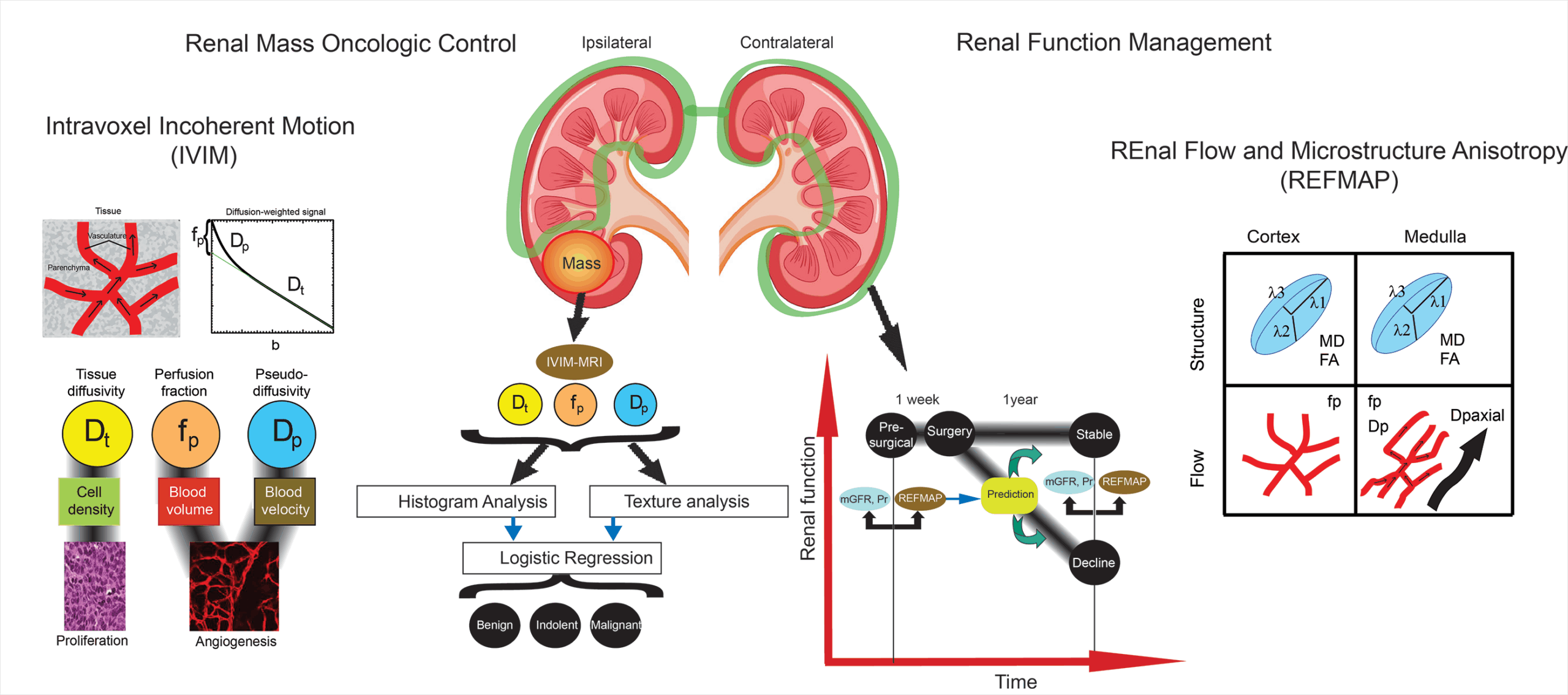
In renal cancer, the primary malignancy and the loss of renal function after partial or total nephrectomy cause severe morbidity and mortality. Therefore, methods to noninvasively monitor renal function and predict its robustness against this decline are in high demand. Here we investigate whether a comprehensive approach we call REFMAP, which includes intravoxel incoherent motion (IVIM) and diffusion tensor imaging (DTI) techniques, can fill this need when added to cancer patients’ standard clinical MR workup.

REFMAP jointly collects IVIM and DTI contrasts: IVIM to separate microstructure from microcirculation, and DTI to assess microscopic anisotropy. The resulting dataset is used to classify aggressiveness of the primary renal lesion and to assess and predict renal function after a partial nephrectomy. Our investigation includes a study of renal masses and a study of renal function.

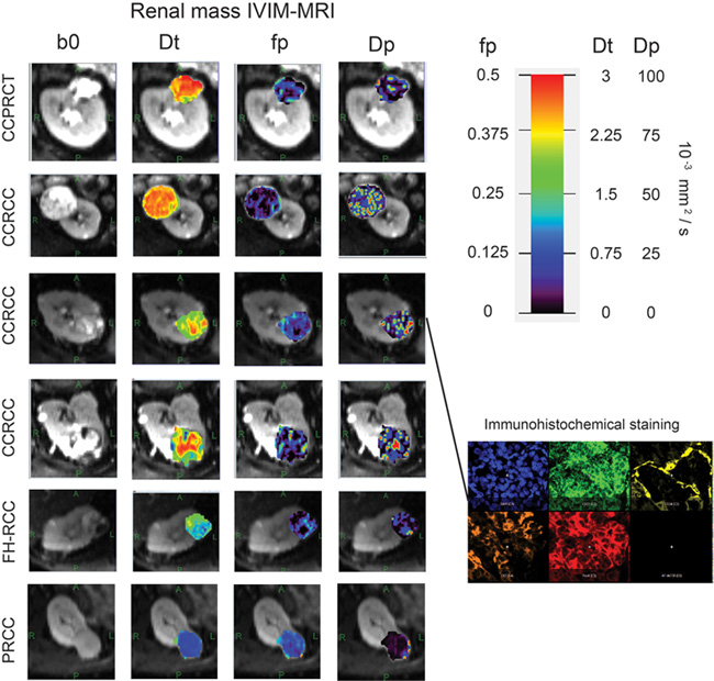
In the renal mass study, we test the diagnostic utility of IVIM biomarkers for discriminating among benign, indolent, and malignant renal masses in a cohort of pre-surgical patients. Using histopathology from the excised tumor as a gold standard, we validate the IVIM metrics and their spatial distribution with quantitative histologic analysis of cell density, microvessel density, and other aspects of the tumor microenvironment.

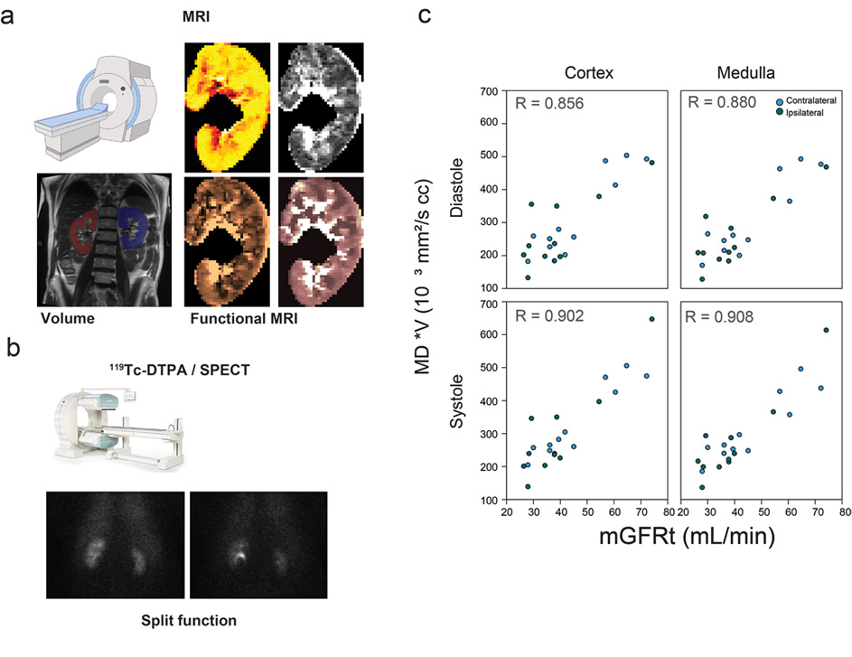
In the renal function study, we determine the potential of REFMAP parameters as a noninvasive surrogate of measured glomerular filtration rate collected in the same patients. We also test the prognostic potential of REFMAP-MRI parameters for predicting renal function response to surgery. These analyses include bilateral MRI kidney sampling, in comparison with split renal function, allowing us to assess compensatory changes between kidneys ipsilateral and contralateral to the lesion.

Keywords
  • Diffusion MRI
  • Renal Cancer
  • Renal Function

Figure 1. IVIM-MRI maps (tissue diffusivity Dt, perfusion fraction fp, and pseudodiffusivity Dp) in renal masses of varying subtypes.  Example immunohistochemical staining of excised specimen is shown illustrating a variety of features (cellularity, microvessel density, etc.)

Figure 2. (a) Functional and anatomic MRI of kidney in renal mass patient. (b) Tc-DTPA tracer clearance imaging in renal mass patient. (c) Correlation of bilateral kidney functional metric (e.g. volume-weighted mean diffusivity) with measured glomerular filtration rate (mGFR) in different tissue and cardiac phase contexts.

Project Team
External Collaborators

Publications

  1. Gilani N, Mikheev A, Brinkmann IM, et al. Spatial profiling of in vivo diffusion-weighted MRI parameters in the healthy human kidney. MAGMA. 2024;37(4):671-680. doi:10.1007/s10334-024-01159-6 
  2. Gilani N, Mikheev A, Brinkmann IM, et al. Characterization of motion dependent magnetic field inhomogeneity for DWI in the kidneys. Magn Reson Imaging. 2023;100:93-101. doi:10.1016/j.mri.2023.03.008
  3. Sigmund EE, Mikheev A, Brinkmann IM, et al. Cardiac Phase and Flow Compensation Effects on REnal Flow and Microstructure AnisotroPy MRI in Healthy Human Kidney. J Magn Reson Imaging. 2023;58(1):210-220. doi:10.1002/jmri.28517
  4. Stabinska J, Wittsack HJ, Lerman LO, Ljimani A, Sigmund EE. Probing Renal Microstructure and Function with Advanced Diffusion MRI: Concepts, Applications, Challenges, and Future Directions. J Magn Reson Imaging. 2024;60(4):1259-1277. doi:10.1002/jmri.29127
  5. Gaing B, Sigmund EE, Huang WC, et al. Subtype differentiation of renal tumors using voxel-based histogram analysis of intravoxel incoherent motion parameters. Invest Radiol. 2015;50(3):144-152. doi:10.1097/RLI.0000000000000111
  6. Notohamiprodjo M, Chandarana H, Mikheev A, et al. Combined intravoxel incoherent motion and diffusion tensor imaging of renal diffusion and flow anisotropy. Magn Reson Med. 2015;73(4):1526-1532. doi:10.1002/mrm.25245

Acknowledgements

We acknowledge support from the following NIH grants: R01CA245671, P41EB017183.