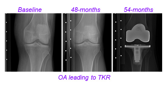
Projects

  • All
  • Aging
  • AI
  • Brain
  • Breast
  • Coils
  • Diffusion MRI
  • Kidneys
  • Molecular Imaging
  • MSK
  • Pulse Sequences
  • Quantitative MRI
  • Simulations
  • Software
  • X-nuclei

Multinuclear MRI to Assess Joint Homeostasis after Knee Injury

This study aims to develop a predictive model for post-traumatic osteoarthritis (PTOA) progression following anterior cruciate ligament (ACL) injury by integrating imaging, biological, and biomechanical markers to improve understanding, therapeutic targeting, and treatment monitoring.

Multinuclear MRI to Monitor Breast Cancer Therapy

This project develops a multinuclear MRI technique using sodium (23Na) and hydrogen (1H) MR fingerprinting at 3 T to create imaging biomarkers for assessing early breast cancer response to neoadjuvant chemotherapy (NACT) and guiding treatment decisions.

Multinuclear Fingerprinting

This project develops a noninvasive MRI technique called multinuclear fingerprinting (MNF) at 7 T, combining hydrogen (1H) and sodium (23Na) magnetic resonance fingerprinting with a super-resolution algorithm to create multi-parametric structural and metabolic maps of the human brain, offering insights into its morphology, physiology, and disorders.

Multiparametric Mapping of Knee Joint with Magnetic Resonance Fingerprinting

This project develops advanced magnetic resonance fingerprinting (MRF) methods enhanced with machine learning to improve the efficiency and robustness of MRI for early detection of osteoarthritis (OA) in the knee by identifying biochemical and structural changes before visible damage occurs.

Longitudinal Single-Center Study with Rapid Quantitative Assessment of Knee Joint with Compressed Sensing

This study uses compressive sensing (CS) techniques to accelerate knee MRI and track osteoarthritis (OA) progression by measuring T2 and T1rho relaxation times, enabling faster detection of cartilage degeneration and changes associated with advancing disease.

Data-Driven Learning Framework for Fast Quantitative Knee Joint Mapping

This project leverages advanced machine learning and deep learning to optimize T2 and T1rho mapping for knee osteoarthritis (OA), aiming to accelerate MRI techniques and enable earlier detection of cartilage degeneration.

In Vivo Insights into Aging-Related Small-Vessel Changes Using USPIO-Enhanced MRI

This project aims to develop an ultra-high-resolution USPIO-enhanced MRI technique to visualize the brain's microvascular architecture, quantify vascular density, and investigate age-related neurovascular changes, potentially advancing the understanding of microvascular aging and its role in neurologic disorders.

Multi-Scale and Multi-Modality Imaging of Neuropathology in VCID

This study aims to advance understanding of neurodegeneration in VCID by developing postmortem MRI protocols, computational tools, and multi-modal atlases, standardizing MRI and histology methods, and creating extensive imaging and biomaterial resources to support research in AD, ADRD, and related pathologies.

Genomic and Imaging Markers to Understand and Predict Progression of Joint Damage After Injury

This study combines genomic analysis and diffusion tensor imaging to identify predictive biomarkers for the risk of developing post-traumatic osteoarthritis (PTOA) following anterior cruciate ligament (ACL) injury in young adults, aiming to improve prevention and therapy development.

Imaging and Therapeutic Targeting of Tissue Crosstalk in the Injured Knee

This project investigates the interplay between inflammation and apoptosis in post-traumatic osteoarthritis (PTOA) using in vitro and in vivo methods to develop therapeutic strategies that target both pathways and prevent disease progression.

Fingerprinting-Based Neuronal Fiber Identification in Brain Surgery

This study aims to advance understanding of neurodegeneration in VCID by developing postmortem MRI protocols, computational tools, and multi-modal atlases, standardizing MRI and histology methods, and creating extensive imaging and biomaterial resources to support research in AD, ADRD, and related pathologies.

FireVoxel: Interactive Software for Multi-Modality Analysis of Dynamic Medical Images

FireVoxel is a freely distributed medical imaging analysis software developed at NYU Langone that provides powerful quantitative tools—especially for dynamic abdominal and genitourinary studies—and has become widely adopted in MRI research worldwide.

Neuroimaging Core of NYU Langone’s Alzheimer’s Disease Research Center

The neuroimaging core of NYU Langone’s Alzheimer Disease Research Center supports research on Alzheimer’s and related dementias by developing and applying advanced imaging techniques to aid early diagnosis, track disease progression, and inform novel diagnostics and treatments.

Breast cancer Intravoxel Incoherent Motion Multisite (BRIMM) Project

The BRIMM project is a multi-site breast cancer study using intravoxel incoherent motion (IVIM) MRI to develop a robust, cross-platform imaging biomarker that noninvasively characterizes the tumor microenvironment and predicts response to neoadjuvant chemotherapy.

Cloud MR: An Open-Source Software Framework to Democratize MRI Training and Research

This project aims to democratize MRI research by developing Cloud MR, an open-source virtual scanner platform accessible via web browser, enabling MRI experimentation, education, and development without the need for physical MRI machines.

Advanced Diffusion MRI in Renal Cancer: Oncologic Control and Renal Functional Reserve

This project investigates REFMAP, an advanced diffusion MRI approach combining IVIM and DTI, to noninvasively diagnose kidney tumors, estimate glomerular filtration rate, and predict renal function outcomes following surgery.

Noninvasive Estimation of Electrical Properties from Magnetic Resonance Measurements

This project develops a three-pronged strategy—combining global inverse modeling, physics-informed neural networks, and vision transformers—to generate high-resolution maps of tissue electrical properties using MRI, with broad applications in medical imaging and treatment.

Open-Source Software Tools for Rapid Radiofrequency Coil Modeling and Simulation in MRI

This project develops open-source, memory-efficient electromagnetic simulation tools using tensor decompositions and optimization techniques to improve RF coil modeling for ultra-high field MRI, enhancing image quality and safety while reducing computational and licensing barriers.