In this project, we investigate, develop, and extend GRASP, a method that enables fast, motion-robust, dynamic MRI with a growing roster of applications and a yearslong record of clinical translation.
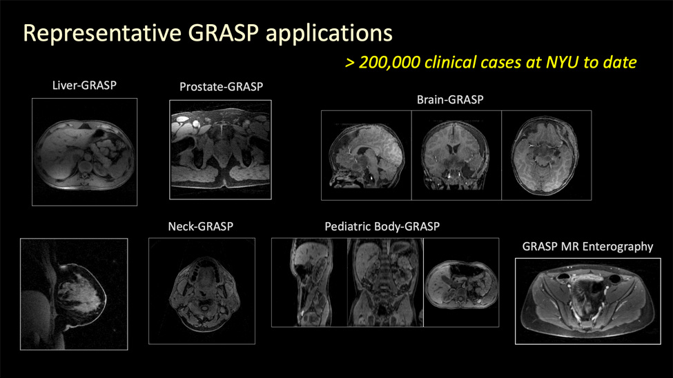
GRASP MRI is one of the signature projects at NYU Langone’s Center for Biomedical Imaging and has been under continuous development for more than a decade. It is a technique that combines a golden-angle radial sampling scheme, compressed sensing, and parallel imaging to enable rapid, motion-robust dynamic MRI. It’s clinical applications span brain, head-and-neck, breast, cardiovascular, pulmonary, renal, abdominal, and pelvic imaging—for both qualitative and quantitative assessment.
GRASP MRI has been fully integrated into the clinical radiology workflow at NYU Langone Health since 2013, with over 200,000 clinical scans performed to date. It is also available on the latest Siemens scanners under the name “GRASP-VIBE” and has been disseminated to hospitals worldwide with widely recognized clinical impact.
Since its initial development, GRASP MRI has evolved into multiple advanced versions – such as XD-GRASP, RACER-GRASP, Dixon-GRASP, GRASP-Pro, and MP-GRASP, each of which further improve imaging performance and expand clinical applications. In recent years, GRASP MRI has also been extended beyond diagnosis toward therapeutic guidance. In particular, the use of XD-GRASP for motion-resolved sparse 4D MRI has shown increasing value in MRI-guided radiation therapy. Recently, a deep learning version of GRASP, called DeepGrasp, has been proposed to address major limitations in the traditional MRI reconstruction pipeline.
Several GRASP software packages are available to MRI researchers interested in implementing this technique, and they can be found among the resources offered on the website of NYU Langone’s Center for Advanced Imaging Innovation and Research. These tools have seen increasing use within the research community and have supported many studies in both technical development and clinical application.