Longitudinal Single-Center Study with Rapid Quantitative Assessment of Knee Joint with Compressed Sensing

Overview
In this study we use our compressive sensing (CS) techniques to accelerate magnetic resonance imaging (MRI) of the knee and track progression of osteoarthritis (OA).
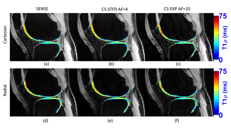
Knee OA is a common degenerative joint disease that causes breakdown of cartilage and leads to biochemical, structural, and morphological changes. There is currently no cure for OA. Early detection of cartilage degeneration is crucial to intervention and drug research, and requires in vivo detection of changes before visible damage occurs. Quantitative MRI can be used to evaluate articular tissue by measuring T2 and T1rho relaxation times but scans are slow. CS techniques can shorten MRI acquisitions and be used to obtain T2 and T1rho maps more quickly.
In the study, people with knee OA and healthy controls receive a baseline MRI, and those with OA receive a follow-up imaging exam two years later. The scans are conducted with our recently developed CS methods. Changes in relaxation times can reveal which participants have OA that is progressing to a more advanced stage.
Keywords
- Knee OA
- T1rho
- Compressed Sensing
Project Team
External Collaborators
- Gregory Chang, MD, MBA , NYU Langone Health
- Steven Abramson, MD, NYU Langone Health
- Jonathan Samuels, MD, NYU Langone Health
- Ricardo Otazo, PhD, MSc, BSc, Memorial Sloan Kettering Cancer Center
Publications
- Zibetti MVW, Sharafi A, Otazo R, Regatte RR. Accelerated mono- and biexponential 3D-T1ρ relaxation mapping of knee cartilage using golden angle radial acquisitions and compressed sensing. Magn Reson Med. 2020;83(4):1291-1309. doi:10.1002/mrm.28019
- Zibetti MVW, Helou ES, Regatte RR, Herman GT. Monotone FISTA with Variable Acceleration for Compressed Sensing Magnetic Resonance Imaging. IEEE Trans Comput Imaging. 2019;5(1):109-119. doi:10.1109/TCI.2018.2882681
Acknowledgements
We acknowledge support from the following NIH grant: NIH R01 AR076328.


