This project focuses on the development of magnetic resonance fingerprinting (MRF) methods for magnetic resonance imaging (MRI) of the knee in order to improve early detection of osteoarthritis (OA).
Knee OA is a common degenerative joint disease that causes breakdown of knee cartilage and leads to biochemical, structural, and morphological changes. There is currently no cure for OA. Early detection of cartilage degeneration, critical for interventions and research, requires identifying changes in vivo before visible damage occurs. Quantitative MRI metrics of T1, T2, and T1rho relaxation can detect changes in articular extracellular matrix. However, existing methods are slow and sensitive to noise and magnetic field variations.
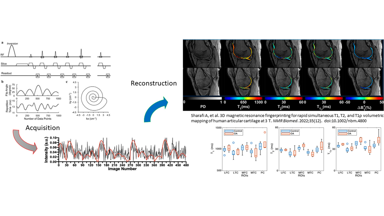
MRF is a relatively new technique that makes use of dynamic signal patterns and is capable of simultaneously estimating multiple MRI parameters, an approach that makes scans more efficient and more robust for musculoskeletal applications. In this project, we enhance MRF with advanced machine learning techniques to improve early detection of OA in the knee.