In this project we pursue a three-pronged research strategy to obtain accurate maps of the electrical properties of biological tissue.
The electrical properties (EP) of biological tissue, namely electric conductivity and relative permittivity, provide quantitative information that has the potential to advance technical, medical, and scientific applications of MRI. These include: more precise transmission and detection of radiofrequency (RF) waves; provision of insights needed to solve the problem of magnetic field inhomogeneity; personalized estimation of RF power specific absorption rate (SAR); better detection of cancer; improved RF ablation and hyperthermia; and discovery of new knowledge about the structure and function of living tissue. However, despite decades of research, EP tissue mapping remains an unsolved problem.
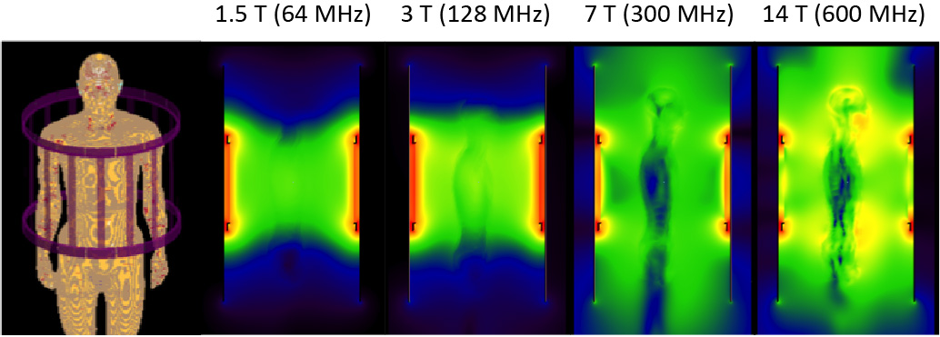
Magnetic resonance, which can provide tomographic measurements that reflect the curvature of the electromagnetic field inside an object, is a promising tool for robust EP estimation. Our goal is to use previously untapped properties of RF coils already integral in MRI to measure electrical properties of tissue at high spatial resolution.
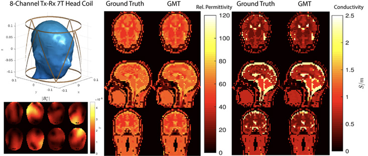
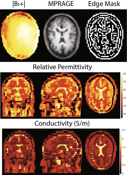
Our strategy comprises three different approaches. First, we solve a global inverse problem by using the integral form of Maxwell’s equations. To this end, we have introduced global Maxwell tomography (GMT), a technique for three-dimensional noninvasive EP estimation from MR measurements. Second, we implement a physics-informed neural network that solves the differential form of Maxwell’s equations. Our method, called physics-informed Fourier networks for electrical properties tomography (PIFON-EPT), estimates EP and magnetic transmit field distributions from noisy or incomplete MR measurements. Third, we have developed a supervised neural network based on a 3D vision transformer to reconstruct EP from MR measurements obtained with a birdcage coil. We are pursuing further development of these techniques with the aim of creating spatially-resolved maps of the electrical conductivity and permittivity of tissue in vivo.