In this project, we are developing translation-ready quantitative MRI methods for early diagnosis and evaluation of femoroacetabular impingement (FAI).
FAI is a pathology of the hip joint in which bony abnormalities cause repetitive contact between the acetabular rim and the proximal femur, eventually leading to hip osteoarthritis. Restoration of pain-free functionality and prevention or delay of end-stage osteoarthritis are in some cases possible with surgery, but success depends on early diagnosis, which can be difficult. Although quantitative radiologic measures can aid in the diagnosis and characterization of FAI, current imaging methods have limited reliability and standard MRI can only detect macroscopic changes associated with advanced lesions. In this investigation, we are developing clinically-viable methods to obtain MRI parameters that can inform early assessment of FAI and help determine which patients are likely to benefit from surgery. We are using model-based quantitative MR techniques capable of rapid, high-resolution, simultaneous mapping of multiple parameters in the hip cartilage with a single scan. Our research on quantitative assessment of cartilage with a combination of model-based parameter estimation, automatic slice positioning, and semiautomatic segmentation has shown potential for high reproducibility. We aim to translate such assessments into routine hip imaging protocols, then use them in longitudinal studies and multicenter clinical trials.
We are also exploring the possibility of fully automated diagnosis of FAI with radiomics, an alternative to the manual or semi‐manual extraction of radiologic measures from radiographs and cross‐sectional imaging. To this end, we have developed a pipeline that automatically segments the hip bones on MR images and extracts quantitative features in order to predict and characterize FAI. Our studies have demonstrated that our proposed radiomics approach can detect subtle shapes and textures on MR images and achieve exceptional performance compared to the standard radiologic measures currently available in the clinic.
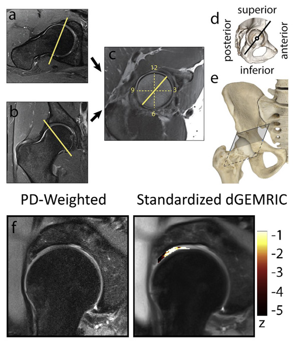
Figure 1. Standardized dGEMRIC analysis in FAI. A clock face image (c) was obtained from true axial (a) and coronal views (b) of the hip. A morphologic MR image and a quantitative dGEMRIC map were acquired for a radial section at the 1:30 clock location, corresponding to the anterior-superior (AS) region of the hip cartilage (d, e). A standardized dGEMRIC map was generated using the central femoral cartilage as the healthy reference. The PD-weighted radial image is shown next to a corresponding Standardized dGEMRIC map (f), where the spatial distribution of the z values in the weight-bearing portion of the hip articular cartilage is superimposed to an MR image of the hip. Values smaller than zero indicate GAG concentration lower than normal. Cartilage was found to be injured during arthroscopy and it clearly shows as damaged (i.e., z < -2) on the Standardized dGEMRIC map, whereas it was ruled as normal by radiologic assessment based on the PD-weighted image.
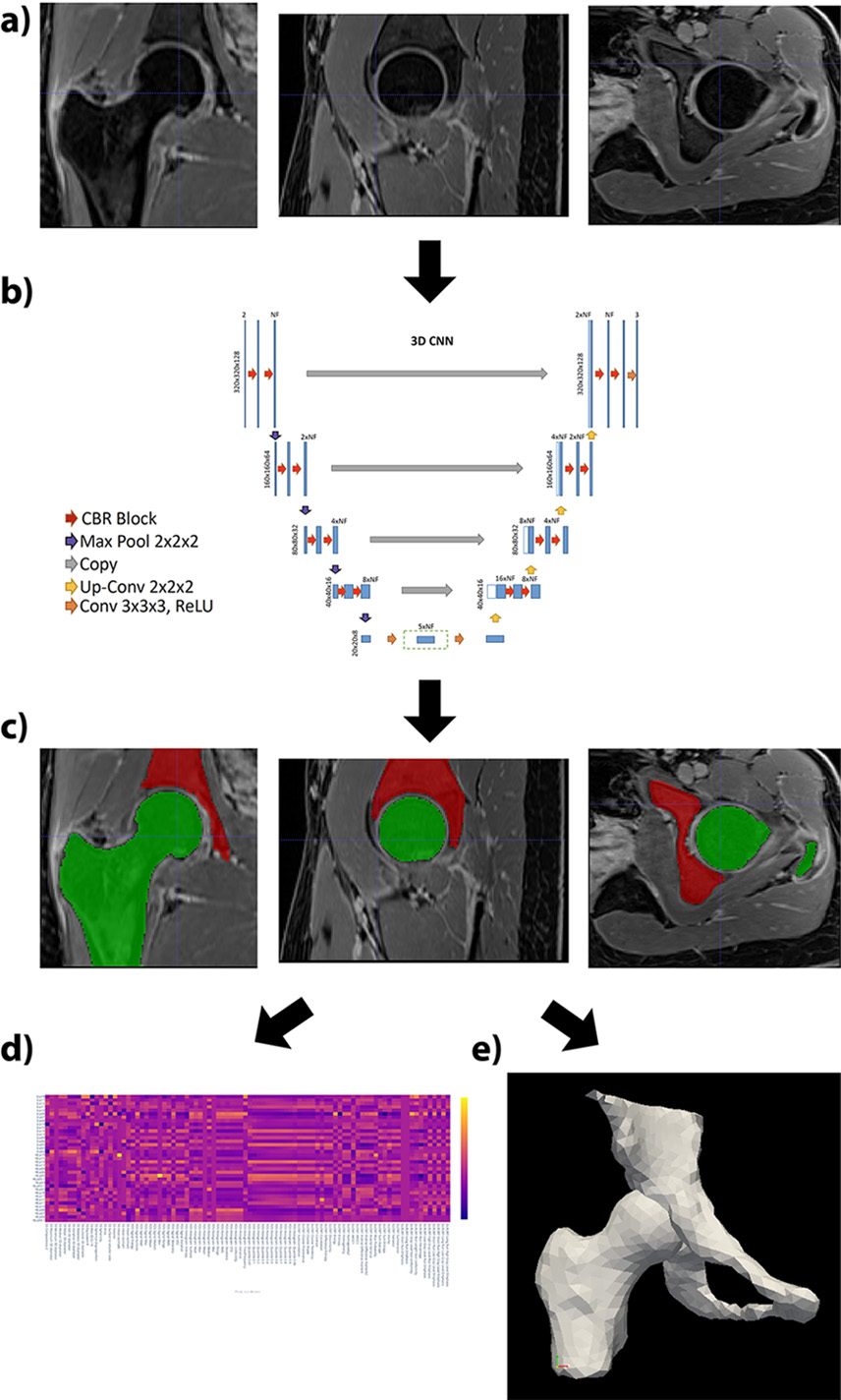
Figure 2. Automatic processing pipeline for the analysis of radiomics features and hip bone morphology in FAI. Coronal, sagittal and axial views of the hip joint were obtained from a water-only image of a 3D Dixon MRI data set (a). A 3D convolutional neural network (b) was used for the automatic segmentation of the acetabulum and the femur (c). The segmentation masks were then used to extract radiomics features (d) and reconstruct a 3D model of the hip joint (e).
Project Lead
This research project has been partially supported by National Institutes of Health (NIH) under Award Number R01AR070297 and R21EB020096.
Researchers at the Center for Biomedical Imaging at NYU Langone Health develop transformative imaging technologies to advance basic science and address unsolved clinical problems.
660 First Avenue
4th Floor
New York, NY 10016