This study combines imaging, biological, and biomechanical markers to develop a predictive model for the progression of post-traumatic osteoarthritis (PTOA) after injury to the anterior cruciate ligament (ACL).
PTOA often occurs after an ACL rupture regardless of how the ACL injury is treated. This suggests that the cellular and molecular changes that directly follow an ACL injury contribute to the development of PTOA.
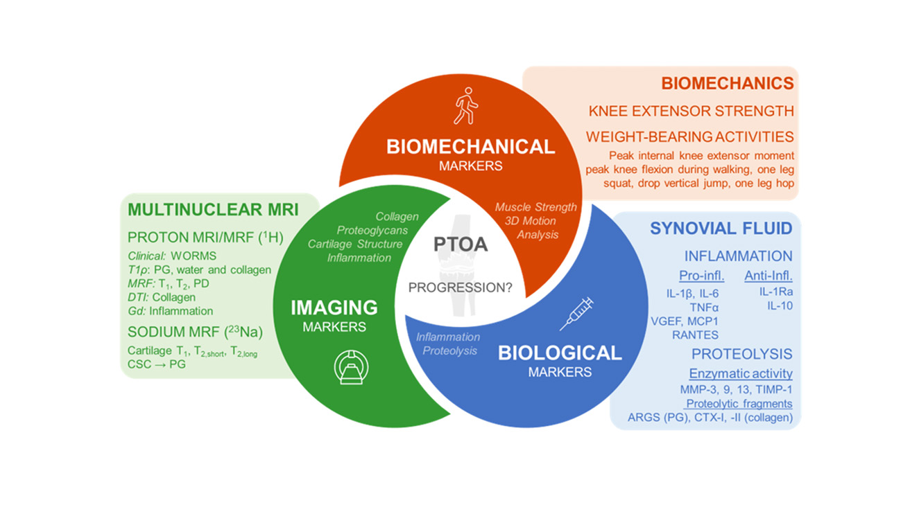
The goal of this study is to develop a predictive model of progression to PTOA by using a combination of signals: multinuclear magnetic resonance imaging (MRI) markers from proton (1H) and sodium (23Na) imaging at 3 T and 7 T; soluble synovial fluid biological markers; and biomechanical markers acquired from patients just after ACL injury and at different times after joint repair. This prognostic combination of biomarkers has the potential to help identify therapeutic targets and monitor the efficacy of intervention in the development of treatments for PTOA.