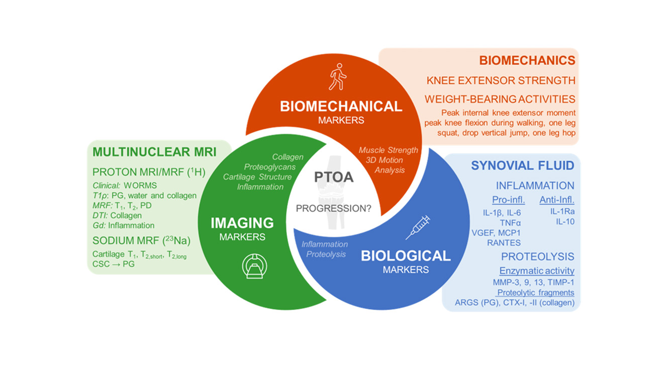
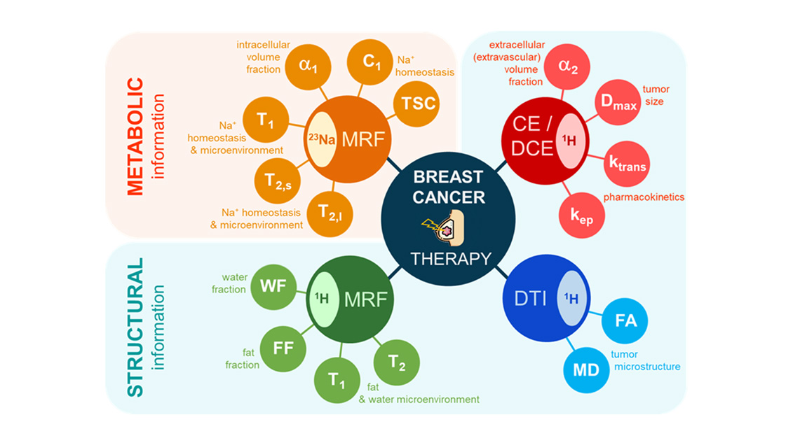
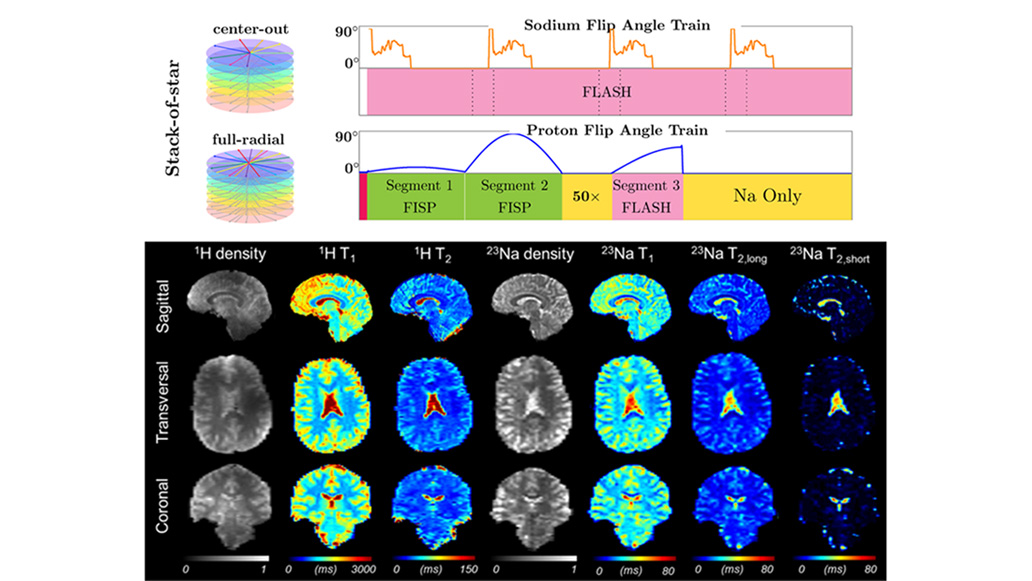
I am developing methodologies for quantitative metabolic X-nuclei magnetic resonance imaging (MRI) in humans in vivo.
X-nuclei MRI is based on the detection of nuclei other than the nucleus of hydrogen (or proton, 1H) from water molecules in the body, as is used in standard MRI. MRI of the sodium (23Na), phosphorus (31P), deuterium (2H), or oxygen (17O) nuclei, for example, can provide brand new metabolic information in tissues as a complement to standard structural 1H MRI. In particular, imaging of the endogenous Na+ ion content and relaxation properties with MRI can help assess fundamental biochemical information related to tissue ionic homeostasis and cellular energetic metabolism, which are not available with other imaging technique in vivo.
Overall, X-nuclei MRI could therefore generate unique imaging biomarkers for detecting early signs of loss of tissue viability due to the disruption of the cellular metabolism in many diseases, but also for monitoring the early effects of therapies targeting different aspects of this cellular metabolism.