Neural Underpinning of Cognitive and Behavioral Deficits in Psychotic Spectrum Disorders

Overview

We are working to uncover the mechanisms of psychotic spectrum disorders and develop biomarkers for gauging disease progression and response to treatment.

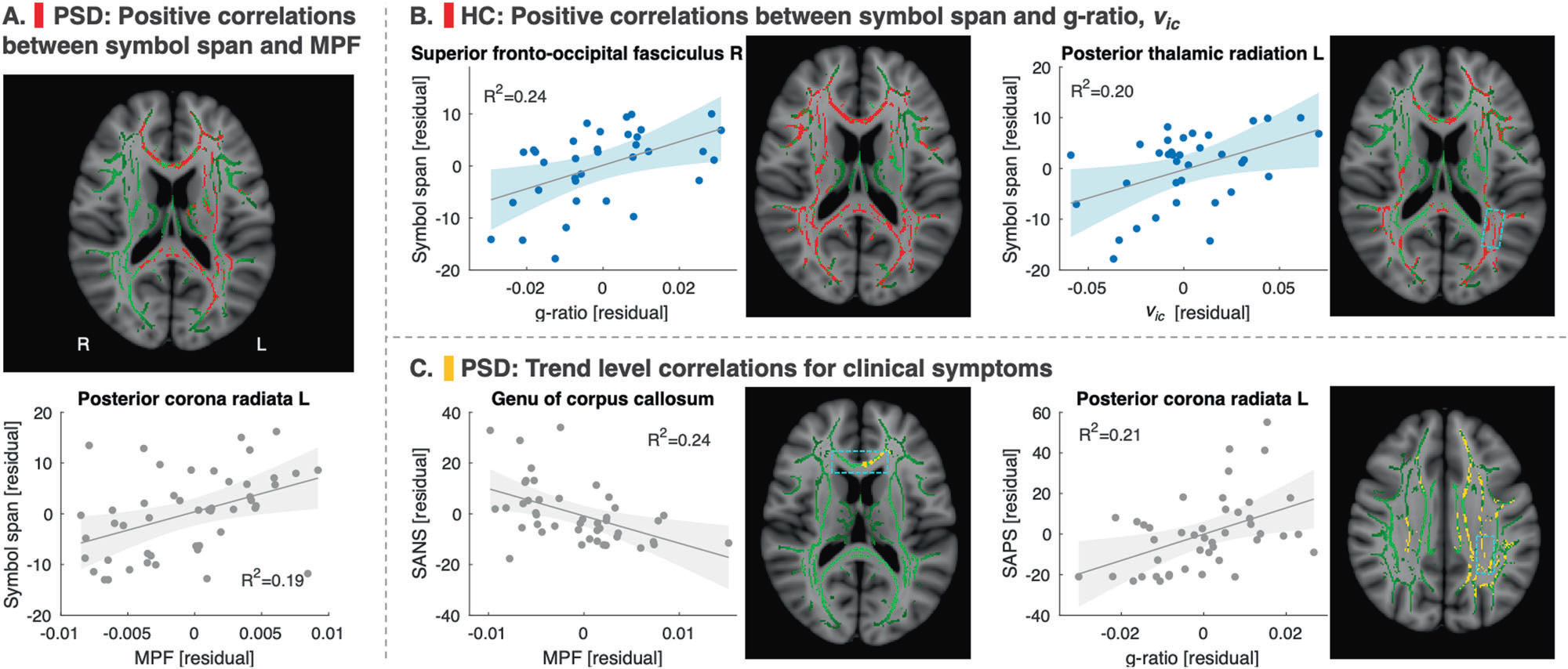
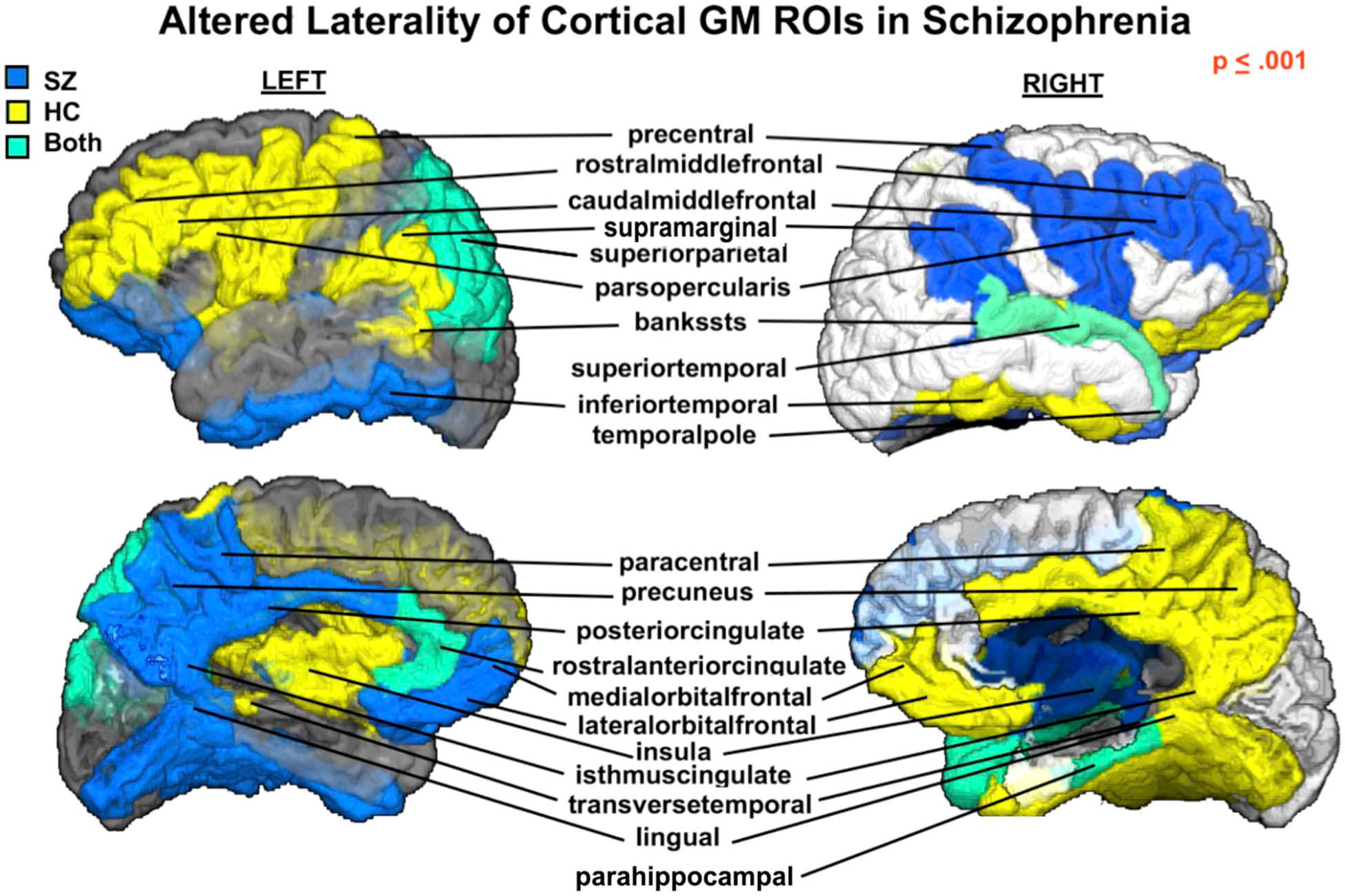
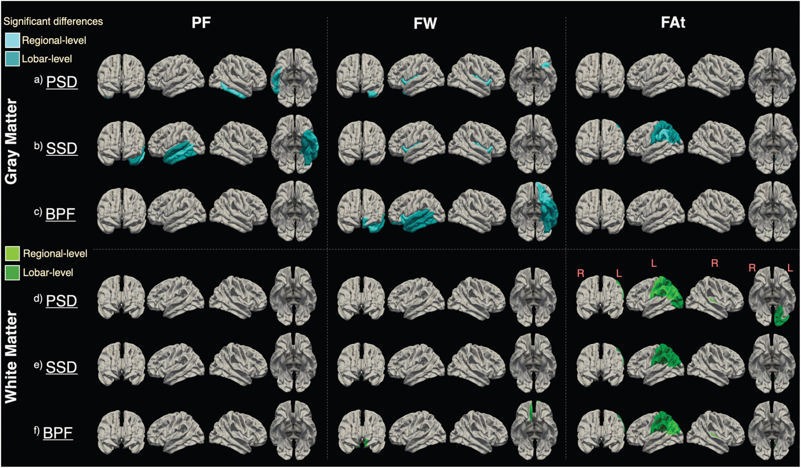
Psychotic Spectrum Disorders (PSD), which include schizophrenia, schizoaffective disorders, and mood disorders with psychotic features, are complex conditions of unclear etiology, variable disease course, and broad range of outcomes. Current medications address only in part the behavioral and cognitive deficits characteristic of PSD. We are pursuing a multi-modal approach focused on 1) revealing the neural mechanisms that underlie the behavioral and cognitive deficits with the goal of informing the development of more efficient therapies and 2) providing objective biomarkers for assessing disease progression and treatment efficacy.  

Keywords
  • Myelin
  • Perfusion
  • Iron
  • Schizophrenia Spectrum Disorders
  • Cognition
Figure 1. Cortical laterality (left vs. right) displayed for regions with significantly different μLI (P ≤ 0.001) in SZ compared with HCs. Color indicates group (SZ—blue, HC—yellow, SZ&HC—green) μLI direction for each ROI. In the HC group, the fronto-parietal motor and language areas appear left lateralized, whereas the orbitofrontal-cingulate-temporal areas are right lateralized. These lateralization patterns are largely lost in SZ with a reversed trend (right vs. left or left vs. right) observed.
Figure 2. Significant analyses of covariance (ANCOVA) group differences in perfusion fraction (PF), free water fraction (FW), and fractional anisotropy of tissue (FAt) in gray matter (blue) and white matter (green), covaried for subjects’ age and sex. Color indicates significant differences in patient groups compared to healthy controls at regional- (light color) or lobar-level (dark color).
Project Team
External Collaborators

Publications

  1. McKenna FF, Miles L, Babb JS, Goff DC, Lazar M. Diffusion kurtosis imaging of gray matter in schizophrenia. Cortex. 2019;121:201-224. doi:10.1016/j.cortex.2019.08.013
  2. McKenna F, Babb J, Miles L, Goff D, Lazar M. Reduced Microstructural Lateralization in Males with Chronic Schizophrenia: A Diffusional Kurtosis Imaging Study. Cereb Cortex. 2020;30(4):2281-2294. doi:10.1093/cercor/bhz239
  3. Sui YV, Bertisch H, Lee HH, Storey P, Babb JS, Goff DC, Samsonov A, Lazar M. Quantitative macromolecular proton fraction mapping reveals altered cortical myelin profile in schizophrenia spectrum disorders. Cereb Cortex Commun. 2021;2:tgab015. doi:10.1093/texcom/tgab015
  4. Sui YV, McKenna F, Bertisch H, et al. Decreased basal ganglia and thalamic iron in early psychotic spectrum disorders are associated with increased psychotic and schizotypal symptoms. Mol Psychiatry. 2022;27(12):5144-5153. doi:10.1038/s41380-022-01740-2
  5. McKenna F, Gupta PK, Sui YV, et al. Microstructural and Microvascular Alterations in Psychotic Spectrum Disorders: A Three-Compartment Intravoxel Incoherent Imaging and Free Water Model. Schizophr Bull. 2023;49(6):1542-1553. doi:10.1093/schbul/sbad019
  6. Sui YV, Bertisch H, Goff DC, Samsonov A, Lazar M. Quantitative magnetization transfer and g-ratio imaging of white matter myelin in early psychotic spectrum disorders. Mol Psychiatry. 2025;30(6):2739-2747. doi:10.1038/s41380-024-02883-0

Acknowledgements

We acknowledge support from the following grants:R21MH085228, R01MH129934, R01MH108962.