Open-Source Software Tools for Rapid Radiofrequency Coil Modeling and Simulation in MRI

Overview

This project focuses on the development of open-source, rapid, and memory-efficient software tools for radiofrequency (RF) coil modeling in magnetic resonance imaging (MRI), with a focus on ultra-high field (UHF) MRI applications at field strength higher than 3 tesla.

While UHF MRI offers superior anatomical detail for early disease diagnosis, it also introduces challenges due to the shorter RF wavelengths relative to body dimensions. These conditions can lead to complex interference patterns that cause local signal dropouts and RF energy deposition hotspots, which deteriorate image quality and raise patient safety concerns.

To address these issues, we are developing novel electromagnetic (EM) simulation tools based on domain decomposition techniques and integral equation methods (wire, surface, and volume formulations). A key innovation is the integration of tensor decomposition techniques to construct compressed, low-memory representations of EM interactions, thus enabling significant acceleration of simulations. Additionally, we are developing an RF circuit co-simulator that automatizes coil tuning, matching, decoupling, and detuning via a pipeline of particle swarm optimizations.

Our simulation tools are the first to leverage tensor decompositions in RF coil design for MRI. By removing licensing costs and substantially reducing computation times, our software aims to offer accessible, high-performance tools for researchers and engineers developing next-generation MRI hardware.

Keywords
  • Computational Electromagnetics
  • Radiofrequency Coils
  • Numerical Linear Algebra

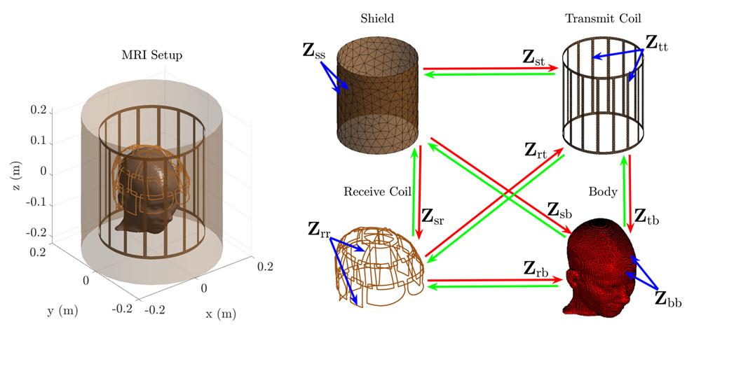
Figure 1. Left: A realistic MRI setup with a 16-leg shielded-birdcage transmitter and a 32-channel receiver. The coils are loaded with a realistic head model. Right: Discretized versions of the shield, transmitter coil, receiver coil, and body model along with a graphical representation of their corresponding self and coupling electromagnetic interactions.

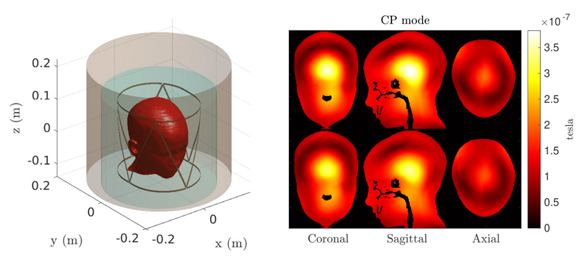
Figure 2. Left: Shielded 8-element triangular coil array loaded with a realistic human head model. The electromagnetic (EM) interactions of the body with conductive elements inside and outside the cyan domain are assembled using the pre-corrected fast Fourier transform and the cross tensor train, respectively. Right: Magnitude of the magnetic transmit field of the triangular coil array in circular polarized (CP) mode combination. Results are shown for 1 mm voxel isotropic resolution, using piecewise constant (PWC) and piecewise linear (PWL) basis functions (top and bottom, respectively), and for three representative orthogonal slices of the head model. The values are masked outside the head model for an enhanced visualization.

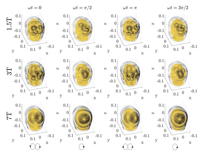
Figure 3. Temporal snapshots of the ideal current patterns on top of a helmet coil former yielding optimal signal-to-noise ratio at a voxel in the back of a realistic head model. The currents are shown for 1.5, 3, and 7 tesla MRI (top to bottom) and for four time-points with equal time differences (left to right).

Project Team
External Collaborators

Publications

  1. Giannakopoulos II, Guryev GD, Serrallés JEC, et al. Compression of volume-surface integral equation matrices via Tucker decomposition for magnetic resonance applications. IEEE Trans Antennas Propag. 2022;70(1):459-471. doi:10.1109/TAP.2021.3090835
  2. Giannakopoulos II, Guryev GD, Serralles JEC, et al. A Hybrid Volume-Surface Integral Equation Method for Rapid Electromagnetic Simulations in MRI. IEEE Trans Biomed Eng. 2023;70(1):105-114. doi:10.1109/TBME.2022.3186235
  3. Giannakopoulos II, Georgakis IP, Sodickson DK, Lattanzi R. Computational methods for the estimation of ideal current patterns in realistic human models. Magn Reson Med. 2024;91(2):760-772. doi:10.1002/mrm.29864

Acknowledgements

We acknowledge support from the following NIH grant: NIH K99EB035163.