My goal is to improve the visualization of the intricacies of the human brain and surrounding cranial nerves. These visualizations aid clinical applications such as neurosurgical planning and help understand and diagnose psychiatric and neurodegenerative diseases. Backed by 16 years of research in (diffusion) MRI physics, diffusion microstructural modeling, and sequence development as well as in the post-processing and interpretation of (diffusion) MRI data in technical and (multi-center) neuroscientific studies, I aim to mine MRI signals, and in particular diffusion MRI signals, for microstructural information affording us insight in central nervous system structure and pathology.
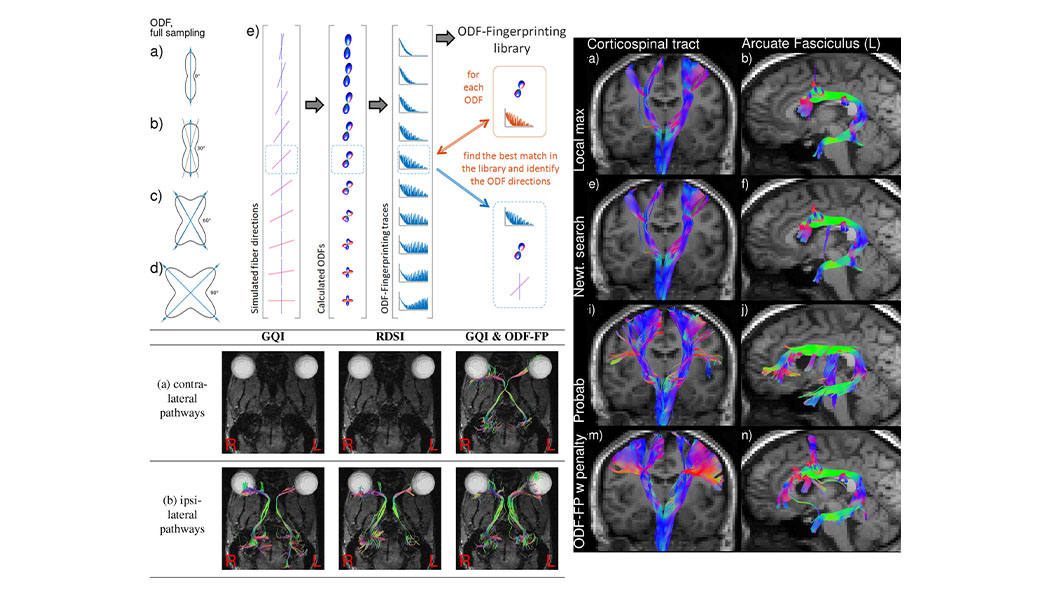
In my recent research, I strive to improve nerve fiber tract visualization and characterization to advance neurosurgical practice. Neurosurgical planning is currently hampered by the failure of fiber tractography methods to reliable reconstruct certain key nerve bundles. By making better use of tissue microstructure information available in diffusion MRI, the ODF-Fingerprinting method increases adherence of visualized fibers to the biological truth. We are also exploring the use of diffusion MRI in evaluating optic nerve inflammation and deterioration for diseases such as glaucoma.
In an equally important aspect of my research, I further strive to evaluate neuroinflammation in the human brain. While TSPO-PET imaging can visualize activated microglia in the brain, inflammatory tissue changes at the microstructural level can also be detected by appropriately modeling the diffusion signal by combining multiple MRI contrasts (diffusion, T2/T2*). My team and I are evaluating neuroinflammation in subtypes of Major Depressive Disorder and in volunteers suffering from Post-Acute Sequelae of COVID-19 where we look at a range of TSPO-PET images, quantitative MRI images and multi-compartment diffusion modeling.