Fingerprinting-Based Neuronal Fiber Identification in Brain Surgery

Overview

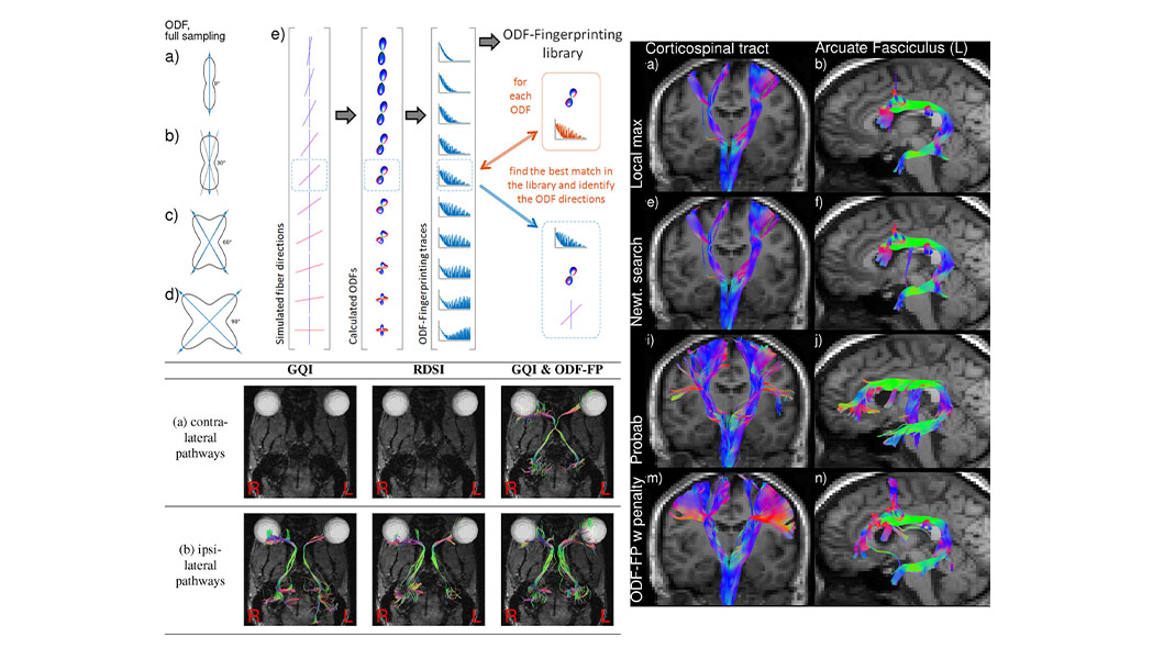
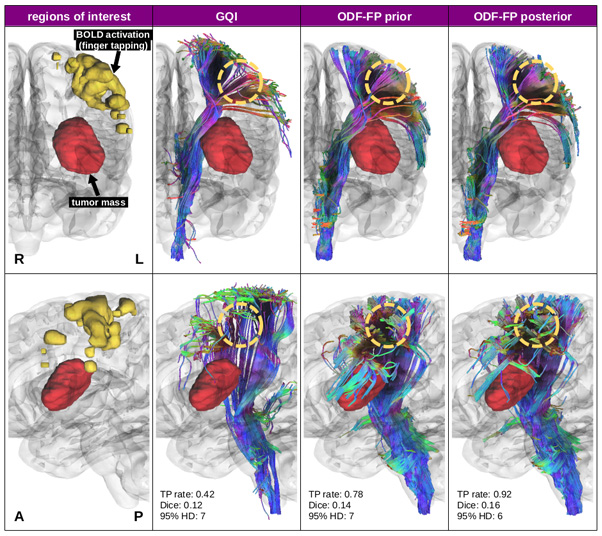
In this project we develop highly precise tractography by using a methodology based on magnetic resonance fingerprinting, which more accurately identifies neural fibers and significantly improves the detection of narrowly crossing fibers.

Diffusion tractography is routinely used to study white matter architecture and brain connectivity in vivo. A key step for successful tractography of neuronal tracts is the correct identification of tract directions in each voxel, but fibers crossing at narrow angles of less than 40 degrees pose challenges to tractography algorithms.

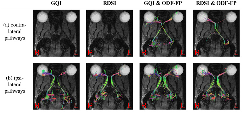
In our method, called ODF-fingerprinting (ODF-FP), fiber configurations are selected based on the similarity between measured orientation distribution functions (ODFs) and elements in a pre-computed library. Unlike traditional approaches that focus only on the ODF maxima, the ODF-FP algorithm relies on the entire ODF shape to infer fiber directions. (In noisy ODFs, the library matching algorithm penalizes the more complex fiber configurations). The technique results in better detection of fiber pairs with small crossing angles while maintaining fiber direction precision, leading to more accurate tractography.

Keywords
  • Neurosurgery
  • Fiber Tractography
  • Diffusion MRI
Figure 1.
Figure 2. Reconstructions of (a) contralateral and (b) ipsilateral optic pathways with generalized q-sampling imaging (GQI), radial diffusion spectrum imaging (RDSI), and ODF-fingerprinting (ODF-FP).
Figure 3. Coronal (top) and sagittal (bottom) views of brain images obtained in a 41-year-old male with high-grade diffuse glioma in the left insula. Yellow circles highlight areas where improved visualization of the lateral projections of the corticospinal tract produced with ODF-FP overlap with the BOLD activation region related to the finger tapping task.
Project Team
External Collaborators

Publications

  1. Filipiak P, Sajitha TA, Shepherd TM, et al. Improved reconstruction of crossing fibers in the mouse optic pathways with orientation distribution function fingerprinting. Magn Reson Med. 2024;91(3):1075-1086. doi:10.1002/mrm.29911
  2. Filipiak P, Shepherd TM, Basler L, et al. Diffusion phantom study of fiber crossings at varied angles reconstructed with ODF-Fingerprinting. Comput Diffus MRI. 2023;14328:23-34. doi:10.1007/978-3-031-47292-3_3
  3. Filipiak P, Shepherd T, Basler L, et al. Stepwise Stochastic Dictionary Adaptation Improves Microstructure Reconstruction with Orientation Distribution Function Fingerprinting. Comput Diffus MRI. 2022;13722:89-100. doi:10.1007/978-3-031-21206-2_8
  4. Filipiak P, Shepherd T, Lin YC, Placantonakis DG, Boada FE, Baete SH. Performance of orientation distribution function-fingerprinting with a biophysical multicompartment diffusion model. Magn Reson Med. 2022;88(1):418-435. doi:10.1002/mrm.29208
  5. Baete SH, Cloos MA, Lin YC, Placantonakis DG, Shepherd T, Boada FE. Fingerprinting Orientation Distribution Functions in diffusion MRI detects smaller crossing angles. Neuroimage. 2019;198:231-241. doi:10.1016/j.neuroimage.2019.05.024

Acknowledgements

We acknowledge support from the following NIH grant: R01EB028774.