Brain Microstructure Imaging

Overview

We are developing and validating biophysical models of diffusion and nuclear magnetic resonance relaxation to quantify the properties of tissue microstructure in the brain.

Our MRI biophysics group develops diffusion methods for estimating and measuring physiological properties at the cellular level of biological tissue, also known as tissue microstructure.

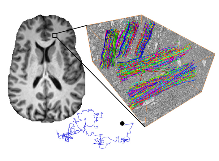
Our team has developed and validated the standard model of diffusion in white matter, a framework for quantifying axonal integrity, inflammation, and fiber orientation dispersion in the brain.1-4 We have also created time-dependent diffusion techniques to quantify axonal diameters, axonal beading and undulations, and water exchange between neurites and extra-cellular space.5-7

To further our research into brain microstructure imaging, we are installing a leading-edge MRI gradient system, Siemens Connectom.X.

Keywords
  • Diffusion MRI
  • Relaxation
  • Microstructure
Project Team
External Collaborators

Publications

  1. Novikov DS, Fieremans E, Jespersen SN, Kiselev VG. Quantifying brain microstructure with diffusion MRI: Theory and parameter estimation. NMR Biomed. 2019;32(4):e3998. doi:10.1002/nbm.3998
  2. Novikov DS, Veraart J, Jelescu IO, Fieremans E. Rotationally-invariant mapping of scalar and orientational metrics of neuronal microstructure with diffusion MRI. Neuroimage. 2018;174:518-538. doi:10.1016/j.neuroimage.2018.03.006
  3. Coelho S, Baete SH, Lemberskiy G, et al. Reproducibility of the Standard Model of diffusion in white matter on clinical MRI systems. Neuroimage. 2022;257:119290. doi:10.1016/j.neuroimage.2022.119290
  4. Coronado-Leija R, Abdollahzadeh A, Lee HH, et al. Volume electron microscopy in injured rat brain validates white matter microstructure metrics from diffusion MRI. Imaging Neurosci (Camb). 2024;2:imag-2-00212. Published 2024 Jul 2. doi:10.1162/imag_a_00212
  5. Novikov DS, Jensen JH, Helpern JA, Fieremans E. Revealing mesoscopic structural universality with diffusion. Proc Natl Acad Sci U S A. 2014;111(14):5088-5093. doi:10.1073/pnas.1316944111
  6. Lee HH, Papaioannou A, Kim SL, Novikov DS, Fieremans E. A time-dependent diffusion MRI signature of axon caliber variations and beading. Commun Biol. 2020;3(1):354. Published 2020 Jul 7. doi:10.1038/s42003-020-1050-x
  7. Jelescu IO, de Skowronski A, Geffroy F, Palombo M, Novikov DS. Neurite Exchange Imaging (NEXI): A minimal model of diffusion in gray matter with inter-compartment water exchange. Neuroimage. 2022;256:119277. doi:10.1016/j.neuroimage.2022.119277

Acknowledgements

We acknowledge support from the following NIH grants: P41EB017183, S10OD034309.