In this project, we are developing a clinically viable MRI technique to create a “microvascular print” of the brain’s blood-vessel architecture; establishing quantitative measures of vascular and capillary density of cerebral structures; and evaluating age-related neurovascular changes in people aged 18 through 85.
Abnormalities in the microvascular arterial system, which delivers the oxygen and glucose that fuel the brain’s metabolism, have been increasingly linked to neurologic disorders, including age-related dementia. However, microvascular aging is itself not well understood, and there is an urgent need for in vivo characterization of this process.
Here, we aim to address this need by creating a way to image arterioles and venules with diameters between 50 μm and 100 μm on 3 T and 7 T MRI scanners, using something we call ultra-high resolution USPIO-enhanced MR arteriogram and venogram (USPIO+MRAV). In developing this new method we are building on our prior work in visualizing small veins and arteries via susceptibility weighted imaging and quantitative susceptibility mapping while modifying the arteries’ magnetic susceptibility with ultra-small-superparamagnetic-iron-oxide (USPIO). If successful, ultra-high resolution USPIO+MRAV has the potential to enable new insights into age-related microvascular changes and to become a significant method in the study of cerebral microvasculature.
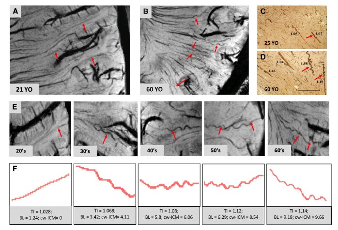
Ferumoxytol-enhanced 3D-GRE MRI significantly improved the detection of cerebral small arteries, enabling the first visualization of tortuous changes in arteries <100μm in diameter. Compared to a 21-year-old (A), a 60-year-old (B) exhibited more tortuous medullary arteries, aligned with Histopathology results (C, D; reprinted from Thore et al., 2007). Artery segments (red arrows) (E) and tortuosity measurements (F) confirmed age-related increases in small artery tortuosity.
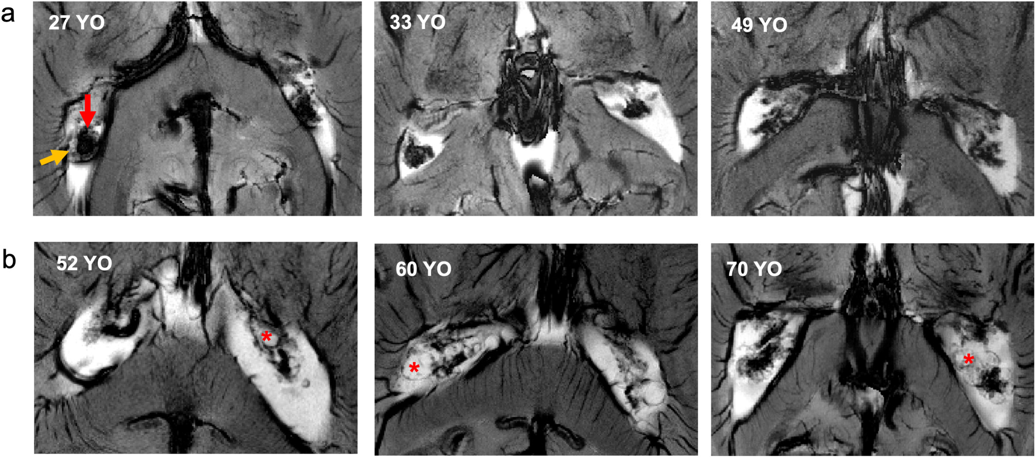
Ferumoxytol-enhanced 2D GRE images of the choroid plexus (ChP) across age groups show: (a) Younger subjects (≤50 years) with densely packed ChP, larger vascular compartments (red arrow), and smaller nonvascular compartments (yellow arrow). (b) Elderly subjects (>50 years) display loosely packed ChP, sparser vascular compartments, and cyst-like degenerative structures (red asterisk).
We acknowledge support from the following NIH grant: NIH R01 NS108491.
Researchers at the Center for Biomedical Imaging at NYU Langone Health develop transformative imaging technologies to advance basic science and address unsolved clinical problems.
660 First Avenue
4th Floor
New York, NY 10016