Magnetic resonance imaging (MRI) provides a highly efficacious means for observing brain anatomy, locating and identifying brain abnormalities, and making diagnosis of brain disorders. Recent advances in state-of-the-art high-field human MR systems have provided ever-advancing imaging capabilities for both clinical diagnosis and basic science research.
Research interests are directed toward developing and applying the quantitative measures at high field MRI in various brain diseases including multiple sclerosis (MS), traumatic brain injury (TBI), brain tumors, and other neurodegenerative diseases. The implementation of these advanced MRI techniques in these diseases has greatly improved our understanding of the dynamics of disease evolution, clinicoradiological correlation, and efficacy of experimental treatments. These advanced techniques include:
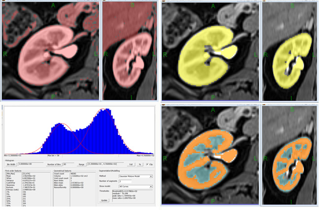
Volumetric and histographical analysis: objective assessment of lesion load, tissue atrophy, and disease activity.
Diffusion tensor imaging (DTI) and fiber tractography (Fig 1): measuring the diffusion characteristics of tissue water can provide information about white matter integrity, connectivity, and pathological substrate of brain lesions.
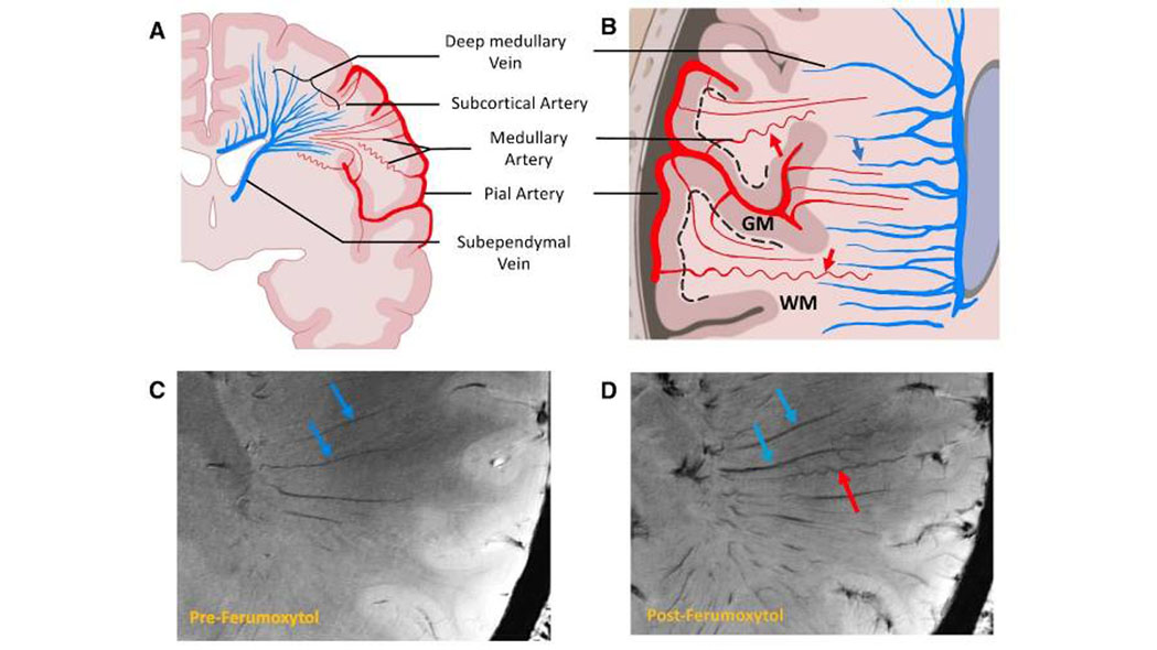
Susceptibility weighted imaging (Fig 2): a 3D high resolution imaging (SWI) that provides high quality of MR venography and susceptibility sensitive technique especially at higher field-strength MR.
MR perfusion and functional MR imaging: mapping cerebral blood flow and volume in tissues to evaluate the critical components of blood supply and hemodynamic conditions in the brain. With event-associated fMRI and resting-state fMRI, brain activity related to a specific task or sensory process as well as functional network can be imaged.
It is believed that at higher field strengths of MR such as 7 Tesla, new areas of research are opening up in microscopic and molecular imaging, biomedical imaging, and functional brain imaging and these ultra-high-field systems will evolve into a new standard for clinical care and scientific discovery.