Tissue Microstructure Imaging in the Body

Overview

We are developing and validating biophysical models of diffusion and nuclear magnetic resonance relaxation to quantify microstructural properties in muscle tissue and the prostate.

Our MRI biophysics group develops diffusion methods for estimating and measuring physiological properties at the cellular level of biological tissue, also known as tissue microstructure.

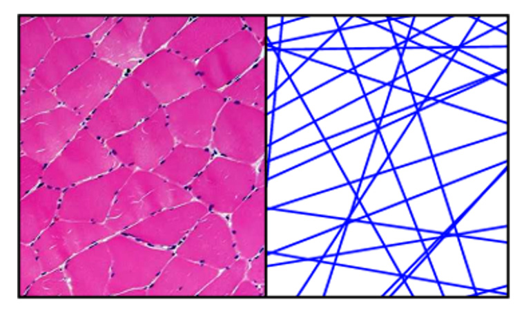
We have pioneered the random permeable barrier model, a framework for quantifying myofiber diameter and membrane permeability in muscle tissue.2-3 Our research on diffusion applications to the prostate includes time-dependent diffusion and diffusion relaxometry acquisitions, which enable mapping fractions of glandular lumen and stroma and lumen sizes.4-5

Keywords
  • Microstructure
  • Muscle
  • Prostate
Project Team
External Collaborators

Publications

  1. Novikov DS, Fieremans E, Jespersen SN, Kiselev VG. Quantifying brain microstructure with diffusion MRI: Theory and parameter estimation. NMR Biomed. 2019;32(4):e3998. doi:10.1002/nbm.3998
  2. Novikov DS, Fieremans E, Jensen JH, Helpern JA. Random walk with barriers. Nat Phys. 2011;7(6):508-514. doi:10.1038/nphys1936
  3. Fieremans E, Lemberskiy G, Veraart J, Sigmund EE, Gyftopoulos S, Novikov DS. In vivo measurement of membrane permeability and myofiber size in human muscle using time-dependent diffusion tensor imaging and the random permeable barrier model. NMR Biomed. 2017;30(3):10.1002/nbm.3612. doi:10.1002/nbm.3612
  4. Lemberskiy G, Rosenkrantz AB, Veraart J, Taneja SS, Novikov DS, Fieremans E. Time-Dependent Diffusion in Prostate Cancer. Invest Radiol. 2017;52(7):405-411. doi:10.1097/RLI.0000000000000356
  5. Lemberskiy G, Fieremans E, Veraart J, Deng FM, Rosenkrantz AB, Novikov DS. Characterization of prostate microstructure using water diffusion and NMR relaxation. Front Phys. 2018;6:91. doi:10.3389/fphy.2018.00091

Acknowledgements

This work was supported by NIH grant R61AT012270.