Understanding CSF Clearance in Aging and Alzheimer’s Brain Through Dynamic Sodium MRI

Overview

We are using advanced MRI techniques to study the relationship among sleep, brain aging, and the clearance of cerebrospinal fluid in healthy controls and people with Alzheimer’s disease.

Neurological health is maintained in part by the brain’s glymphatic system, which flushes away waste by circulating cerebrospinal fluid (CSF). Alzheimer’s disease—characterized by excessive buildup of toxic amyloid beta protein in the brain—is suspected to be causally linked to disrupted CSF clearance. Recent studies of mice have found that impairing the CSF clearance pathway led to a 70-percent reduction in clearing amyloid beta, and that enhancing CSF flow through sleep led to a 100-percent increase in clearing the protein. But it is unknown whether these impairment and enhancement effects exist in humans and, if so, how they change with aging.

In this investigation, our goal is to determine how age affects CSF production, bulk flow, and drainage in the normal brain; how sleep affects CSF clearance in the normally aging brain; and whether CSF clearance is disrupted in patients with Alzheimer’s.

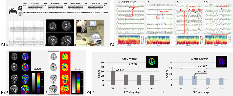
Technical limitations make the study of CSF clearance in humans challenging. Our project addresses these challenges by employing two new methods. First, we acquire dynamic sodium MRI to quantify the velocity of CSF bulk flow in brain parenchyma. Second, using ultrashort echo time (UTE) MRI, we obtain UTE-T2* values to quantify the calcification of the choroid plexus and to assess potential deficiency in the production of CSF. The 0.22-millimeter high resolution of our MR images lets us visualize trabecular structures in arachnoid villi and evaluate the resistance of CSF drainage.

Knowledge about the degeneration of CSF clearance in the normally aging human brain and about the disruption of CSF clearance in the presence of Alzheimer’s has significant potential to help advance both the treatment and the prevention of Alzheimer’s disease.

Keywords
  • Alzheimer’s Disease
  • CSF Clearance
  • Sodium MRI

Figure 1. Sleep study on healthy subjects at 3 T with simultaneous sodium (23Na) MRI and EEG. Panel 1 (P1) shows the study setup, including MRI, EEG, sodium images, and hardware. P2 shows typical EEG waveforms and spectra of sleep stages. P3 shows an example of extracellular volume fraction (ECVF) maps from a  43-year-old female. P4 shows changes in ECVF during sleep stages in both the gray matter (P=0.036) and the white matter (P=0.085, nearly significant).

Figure 2. EEG system testing and preparation. Team members are seen testing our MRI-compatible EEG system (Brain Vision) and preparing on a participant for a sleep study with simultaneous EEG and sodium (23Na) MRI.

Figure 3. Outreach to community. Team members are seen presenting our study to seniors at local community centers during recruitment of healthy volunteers and AD patients.

Videos. Dynamic sodium MRI images (linear interpolation in time for display purpose only) show a continuous flow of CSF into and out of the tissues in the brain for clearance.

a) the 27-year-old male subject

b) the 72-year-old male subject

Project Team
External Collaborators

Publications

  1. Qian Y, Lin YC, Chen X, Ge Y, Lui YW, Boada FE. Single-Quantum Sodium MRI at 3T for the Separation of Mono-and Bi-T2 Sodium Signals. Scientific Reports. June 2025 (accepted). Res Sq [Preprint]. 2025 Apr 7:rs.3.rs-5456028. [Version 1] https://doi.org/10.21203/rs.3.rs-5456028/v1
  2. Alfihed S, Majrashi M, Ansary M, et al. Non-Invasive Brain Sensing Technologies for Modulation of Neurological Disorders. Biosensors (Basel). 2024;14(7):335. Published 2024 Jul 9. doi:10.3390/bios14070335
  3. Watson K, Chen, X, Lin Y-C, Henin S, Kumbella N-M, Quimbo J, Rockowitz Z, Ge Y, Masurkar A, Liu A, Lui YW, Qian Y. Impact of Sleep on Extracellular Volume in the Human Brains: A confirmation study with simultaneous sodium MRI and EEG. In the Proceedings of AAIC25 Annual Meeting. Toronto, Canada. 27–31 July 2025. p.101338. (poster).
  4. Watson K, Chen X, Lin Y-C, Kumbella N-M, Quimbo J, Henin S, Rockowitz Z, Liu A, Masurkar A, Babb J, Ge Y, Lui YW, Qian Y. Impact of Sleep on Extracellular Volume Space in Human Brains: A Simultaneous Study with Sodium MRI and EEG. In the Proceedings of the 33rd ISMRM Annual Meeting. Singapore. 10–15 May, 2025. p.4580. (digital poster).
  5. Lin Y-C, Watson K, Chen X, Henin S, Kumbella N-M, Quimbo J, Ge Y, Masurkar A, Liu A, Lui YW, Qian Y. Simultaneous Dynamic Sodium (23Na) MRI and EEG Analysis of Cerebrospinal Fluid Flow Velocity in the Human Brain During Sleep. In the Proceedings of the 33rd ISMRM Annual Meeting. Honolulu, HI, USA. 10–15 May, 2025. p.5047. (traditional poster).
  6. Das A, Lin Y-C, Qian Y. Optimizing MPPCA for Complex Sodium MRI: Component Analysis and Noise Characterization at 3T. In the Proceedings of the 33rd ISMRM Annual Meeting. Honolulu, HI, USA. 10–15 May, 2025. p.5041. (traditional poster).
  7. Qian Y, Watson K, Chen X, Lin Y-C, Henin S, Kumbella N-M, Quimbo J, Ge Y, Masurkar A, Liu A, Lui YW. Quantum-Sensing MRI: Neuronal Firings in Human Brains under Finger-Tapping in a Wide Range of Ages. In the Proceedings of the 33rd ISMRM Annual Meeting. Honolulu, HI, USA. 10–15 May, 2025. p.3734. (digital poster).

Acknowledgements

This project is supported by the National Institute On Aging (NIA) of the National Institutes of Health (NIH) under Award Number RF1/R01 AG067502.