Multi-Scale and Multi-Modality Imaging of Neuropathology in VCID

Overview

This study aims to advance the understanding of gray and white matter neurodegeneration linked with vascular contributions to cognitive impairment and dementia (VCID) by developing novel post-mortem MRI protocols and computational tools for multi-modal, multi-scale analysis of brain MRI, histopathology, and proteomics.

MRI and histopathology can provide insights into disease mechanisms and uncover therapeutic implications in age-related cognitive impairment and dementia. In this project we are working to standardize MRI and histology protocols, introduce novel tools for postmortem analysis, and provide extensive imaging and biomaterial resources to drive progress in research on VCID in Alzheimer’s disease (AD) and AD-related dementia (ADRD).

In particular, we are pursuing four aims:

  • First, to support integrated MRI and histopathological analysis of AD and ADRD by establishing a postmortem pipeline for brain autopsy, fixation, and sectioning. 
  • Second, to create a multi-modality atlas of the medial temporal lobe and prefrontal cortex with a focus on amyloid-beta-peptide, tau-protein, vascular, and microstructural pathology. The atlas will integrate MRI and pathology data collected by NYU Langone’s Alzheimer’s Disease Research Center from people with AD, ADRD, and traumatic brain injury.
  • Third, to advance the understanding of white matter hyperintensities and small vessel disease through high-resolution voxel-wise mapping of postmortem MRI and histopathology.
  • Last, to develop shared digital libraries of imaging and pathology protocols; software; brain atlases; and data from 75 elderly brains.
Keywords
  • Postmortem MRI
  • Alzheimer’s Disease
  • Histopathology

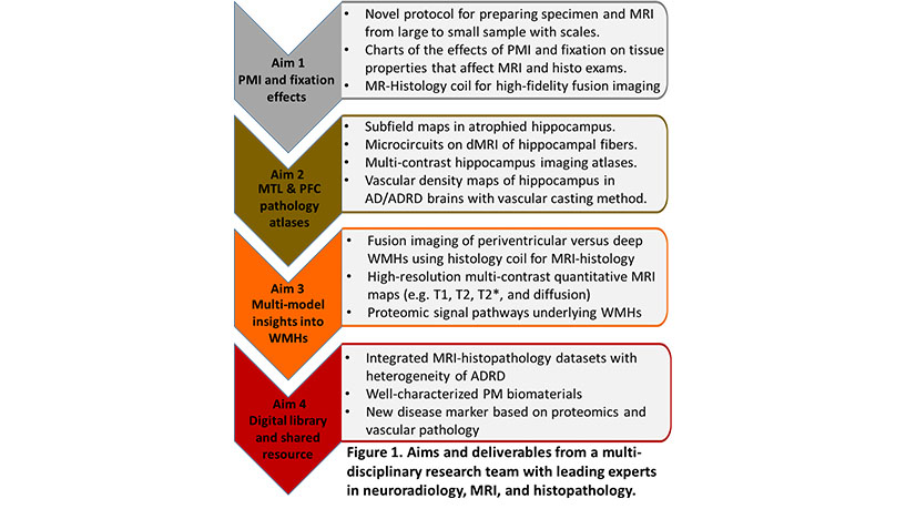
Figure 1. Ex vivo MRI of the hippocampus. (A): MRI co-registered to H&E and SM31-stained images. Major axonal pathways and their orientations can be qualitatively examined. (B): Using hippocampal atlas, pathways between subfields can be reconstructed. Part of the perforant pathway between entorhinal cortex (EC) and dentate gyrus can be reconstructed. (C): Reconstruction of the perforant pathway and Alveus/fimbria, showing the FA values along the perforant pathways, EC volumes, and streamline density.

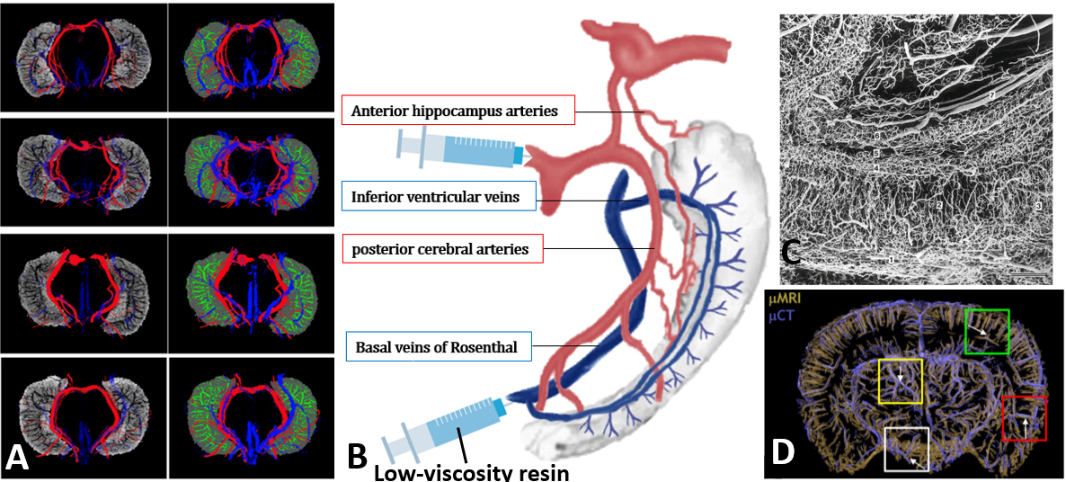
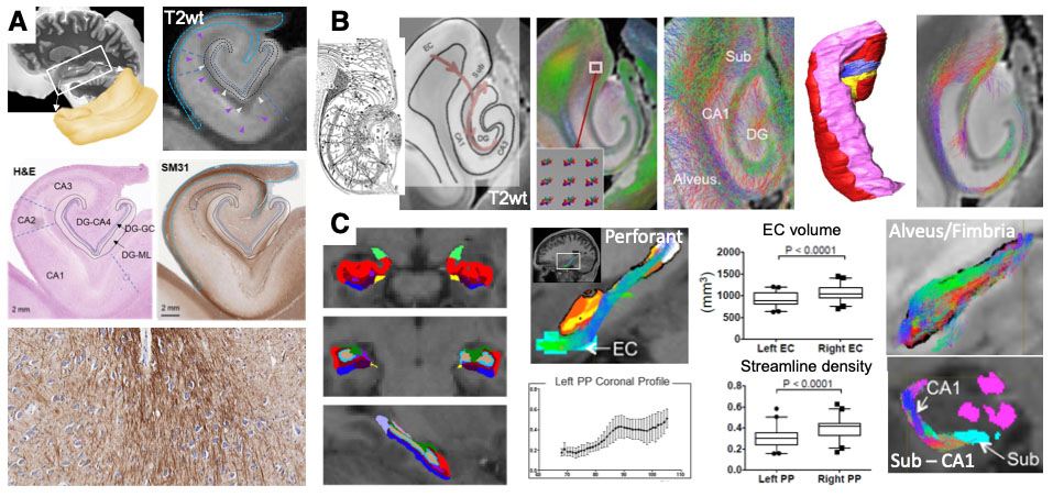
Figure 2. (A) In vivo high-resolution 3D USPIO-enhanced MRAV showed hippocampal vascular map in four healthy volunteers (Buch S et all Neuroimage 2022). (B) Illustration of Microfil vascular casting for hippocampus. (C) Vascular casting results of human hippocampus [Reprint from Duvemoy H, The Human Hippocampus, 2013]. (D) 3D rendering of large blood vessels in a mouse brain based on uMRI (yellow) and uCT (purple).

Figure 3. Multi-parametric MRI probes in an AD patient: Despite WMH lesions in three locations are distinguished on quantitative MRI maps, their underlying pathophysiology is unclear. The proposed MRI-histology strategies (shown on the far right column) will help to fill this knowledge gap.

Project Team
External Collaborators

Publications

  1. Li C, Buch S, Sun Z, et al. In vivo mapping of hippocampal venous vasculature and oxygenation using susceptibility imaging at 7T. Neuroimage. 2024;291:120597. doi:10.1016/j.neuroimage.2024.120597
  2. Sun Z, Li C, Muccio M, et al. Vascular Aging in the Choroid Plexus: A 7T Ultrasmall Superparamagnetic Iron Oxide (USPIO)-MRI Study. J Magn Reson Imaging. 2024;60(6):2564-2575. doi:10.1002/jmri.29381
  3. Sun Z, Li C, Wisniewski TW, Haacke EM, Ge Y. In Vivo Detection of Age-Related Tortuous Cerebral Small Vessels using Ferumoxytol-enhanced 7T MRI. Aging Dis. 2024;15(4):1913-1926. Published 2024 Aug 1. doi:10.14336/AD.2023.1110-1

Acknowledgements

We acknowledge support from the following NIH grant: U24 NS135568.